Name / Period:
Rattus norvegicus 2019
Norway Rat
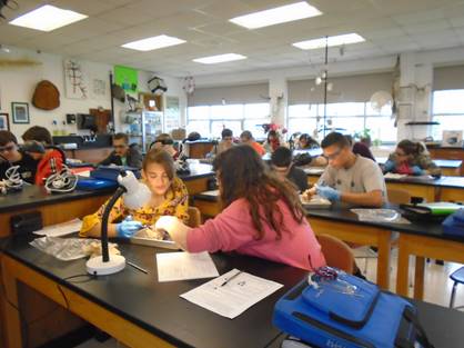
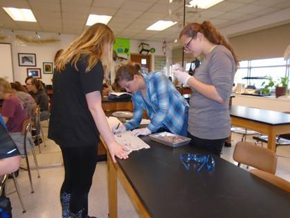
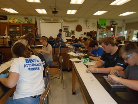
You are to primarily find the following structures and know something about their physiology by using the assigned vocabulary assignment and the lab manual for this investigation. Check each item off as you go along after to have found it on the specimen (not book). Note: “right and left” is based on the specimen’s “Right and Left” The following items may be used for a “lab practical” and/or an “examination” at the end of the lab or year. Care, respect, and professionalism are to be adhered to throughout the lab. You will be graded on how well you stay on task, on identifying the structures, and a lab practical. Be prepared for periodical questioning throughout the activity.
External Features: p5
¾ Pinna
¾ Eyelids
¾ Nares
¾ Anus
¾ * Female urogenital structures: ie vaginal orifice (see p33)
¾ * Male urogenital structures: ie scrotum (see p35)
|
|
|
|
|
|
Skinning the Rat: p4-6
- No need to pin down the specimen.
- Start by creating a small hole in the neck to begin the process.
- Skin is “paper-thin”; pinch/move skin while cutting; do not cut muscle.
Muscular System (Ventral: Front): p8-13
- no need to remove or cut; just identify
¾ Masseter
¾ Pectoralis Major
¾ Pectoralis Minor
¾ Triceps Brachii
¾ Linea Alba (whitish “lined” membrane; not a muscle)
¾ Rectus Abdominis
¾ Gracilis
Muscular System (Dorsal: Back): p15-19
- no need to remove or cut; just identify
¾ Clavotrapezius
¾ Acromiotrapezius (and/or Spinotrapezius)
¾ External Oblique
¾ Biceps Femoris
¾ Lumbodorsal Fascia (whitish/clear membrane on back; not a muscle)
Before Moving On, Ask to Get Permission at This Time / Q&A May Follow
|
|
|
|
|
|
Throat and Oral Cavity: p19-22
¾ Lymph Node(s) ; (3-4) ; (remove clear membrane with probe to see)
¾ Mandibular Gland(s) ; (2) ; (remove clear membrane with probe to see)
¾ Tongue (may not see well, just make note of where the structure is located)
¾ Teeth
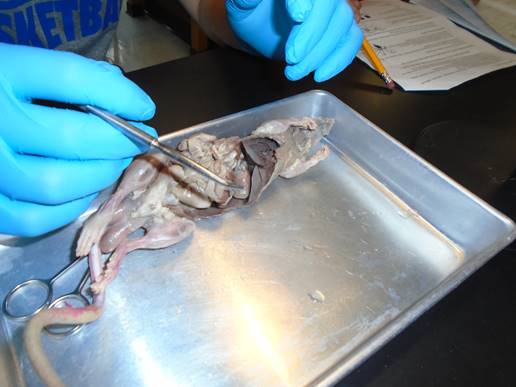
Circulatory System: p24-31
- [Cut] down from the neck to the bottom of the rib cage ; break ribs way back
¾ Thymus Gland (top of the heart; lightly brown/whitish)
¾ Atria (pl.); (r./l.) ; small flaps along the left and right top of the heart
¾ Ventricle(s); (r./l.) [Cut] through horizontally to see (Do Not Remove)]; blue = right ventricle ; red = left ventricle
¾ Spleen (brown, flag-like organ on rat’s left side)
¾ Aortic Arch ; [Push] upwards or remove thymus gland to see
¾ Aorta (behind lungs or intestines) ; red/pinkish
¾ Vena Cava (behind lungs/intestines) ; blue ; next to aorta
¾ Renal Veins and Renal Artery(ies) ; both off the kidneys
Respiratory System: p24-26
¾ Trachea (white cartilaginous ring rings) ; use probe to move brown muscle laying on top of it aside
¾ Lungs (note that there are several lobes (sections) of the lung)
__ Diaphragm (muscle between bottom of lungs and top of the liver) ; you may have cut it in half without knowing
Before Moving On, Ask to Get Permission at This Time / Q&A May Follow
Digestive System: p22-24
- [Cut] down from the rib cage to the tail ; follow the linea alba
- [Cut] the sides laterally towards the backbone to create 1-2 flaps ; do not remove the sides
¾ Liver (made of several “lobes”) ; humans have a “gall bladder” under liver ; unlike rats
¾ Stomach (look under liver ; move thin/clear mesentery away)
¾ Esophagus (best seen by looking under diagram leading into the stomach)
¾ Pancreas (between tubes of the first part of the small intestine ; delicate brownish “granular” looking)
¾ Small Intestine (small in diameter ; not length)
¾ Large Intestine or Colon (large in diameter ; not length)
¾ Peritoneum (clear/thin membrane between intestines)
¾ Cecum ; (beginning of large intestine ; “dead-end” pouch) ; humans have an “appendix” off this ; unlike rats
Urinary System: p32-33
¾ Kidney(s) ; (brown bean shaped organs along the sides ; 2 of them)
¾ Urinary Bladder (small brownish shaped organ)
Reproductive System: p33-35
* pending on your specimen, find someone with the other gender to view their structures (if nobody; use text)
|
Female:
¾ Ovary (Oviducts (yellow coiled tubes)) ¾ Uterus (Uterine Horns) ¾ Vagina |
Male:
¾ Testis
¾ Seminal Vesicles
__ Penis
Before Moving On, Ask to Get Permission at This Time / Q&A May Follow
Central Nervous System: p36-39
¾ Brain [Cut] “around” skull starting through an eye socket; carefully remove top of skull (Do Not Remove brain)
¾ Cerebral Hemisphere of Brain (Cerebrum in humans)
¾ Cerebellar Hemisphere and Vermis Area of Brain (Cerebellum in humans)
¾ Spinal Cord [Cut] vertebra/backbone in half, then bend the specimen in half to see white/pinkish cord cross section