Rattus norvegicus 2015
“Norway Rat”
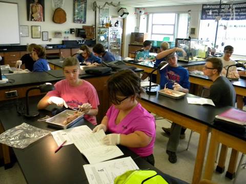
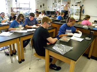
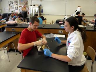
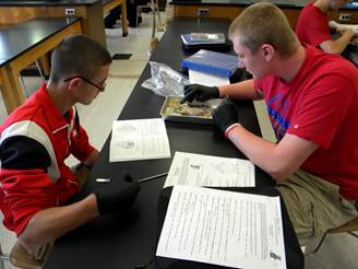
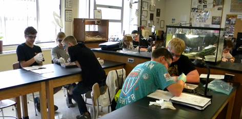
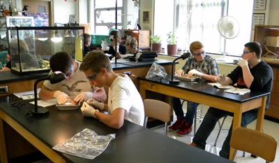
You are to primarily find the following structures and know something about their physiology by using the assigned vocabulary assignment and the lab manual for this investigation. Check each item off as you go along. Note: “right and left” is based on the specimen’s “Right and Left” The following items may be used for a “lab practical” and/or an “examination” at the end of the lab or year. Care, respect, and professionalism is to be adhered to throughout the lab. You will be graded on how well you stay on task and on identifying the structures. Be prepared for periodical questioning throughout the activity.
External Features: p5
¾ Pinna
¾ Eyelids
¾ Nares
¾ Anus
¾ * Female urogenital structures: ie vaginal orifice (see p33)
¾ * Male urogenital structures: ie scrotum (see p35)
* Pending on which gender you have, find someone else in the class who has the opposite and note their anatomy
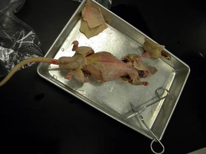
Skinning the Rat: p4-6
- No need to worry about “pinning” specimen.
- If no “hole” in neck, create your own to begin the process.
- Skin is “paper-thin”; be CAREFUL NOT to cut muscle.
Muscular System (Ventral: Front): p8-13
- no need to remove or cut; just identify
¾ Masseter
¾ Pectoralis Major
¾ Pectoralis Minor
¾ Triceps Brachii
¾ Linea Alba (whitish (non-muscle) membrane)
¾ Rectus Abdominis
¾ External Oblique
¾ Gracilis
Muscular System (Dorsal: Back): p15-19
- no need to remove or cut; just identify
¾ Clavotrapezius
¾ Acromiotrapezius (and/or Spinotrapezius)
¾ External Oblique
¾ Triceps Brachii (and/or Biceps Femoris)
¾ Fascia “Lumbodorsal” (whitish (non-muscle) membrane)
Throat and Oral Cavity: p19-22
¾ Lymph Node(s) ; (3-4)
¾ Mandibular Gland(s) ; (2) ; (remove clear membrane to see)
¾ Tongue (may not see well, just make note of where the structure is located)
¾ Teeth
Before Moving On, Ask to Get Permission at This Time / Q&A May Follow
|
|
|
|
|
|
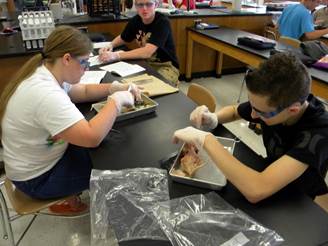
Cutting to Expose the Internal Structures: p22 (may again use p4 as a guide)
Abdominal Cavity and Digestive System: p22-24 (Note: Cut muscle off or aside to view)
¾ Liver (optional: median, left, right, and caudate lobe); gall bladder (stores bile) found in humans, not the rat
¾ Stomach (look under liver) [cut the stomach long-wise and look inside]
¾ Esophagus (note: best seen if you look under trachea (wait to view when you get to the “Respiratory System”)
¾ Pancreas (between small intestine and stomach; within mesentery; delicate, red or brown “granular” looking)
¾ Spleen (brown, flag-like organ on rat’s left side)
¾ Small Intestine (optional: duodenum (beginning), jejunum (middle), ileum (end))
¾ Peritoneum (clear/thin membrane between intestines)
¾ Cecum (or Caecum) ; (between small and large intestine; “dead-end” pouch) ; humans have “appendix” on cecum
¾ Large Intestine (Colon) (optional: ascending (beginning), transverse (middle), descending (end))
Before Moving On, Ask to Get Permission at This Time / Q&A May Follow
Circulatory System: p26-31 (Note: Must cut through sternum and remove ribs to view)
¾ Thymus Gland (top of the heart; lightly brown/whitish)
¾ Pericardium (clear/ thin membrane over heart) ; may have fallen to either side of the heart
¾ Atrium or Atria (pl.); (r./l.)
¾ Ventricle(s); (r./l.) [cut heart long-wise to see them (Do Not Remove)]; blue = right ventricle; red = left ventricle.
Try to Find the Following (If you cannot, that is fine… thanks for trying):
¾ Vena Cava (Anterior (Superior) or Posterior (Inferior))
¾ Aortic “Arch” (Aorta) [cut away thymus gland to see]
¾ Aorta (Thoracic (behind lungs) or Descending (behind intestines)
¾ Renal Veins and Renal Artery(ies) ; (off the kidney)
Respiratory System: p24-26
¾ Trachea (white cartridge ringed structure in throat and down towards lungs) / reminder: find under, the Esophagus
¾ Lung (note that there are lobes (sections of the lung))
__ Diaphragm (muscle between lungs and the liver); (you may have cut it in half without knowing)
Before Moving On, Ask to Get Permission at This Time / Q&A May Follow
Urinary Structures: p32-33
¾ Kidney(s) ; (2)
¾ Urinary Bladder
Female/Male Genital Structures: p33-35
* pending on your specimen, find someone with the other gender to view their structures (if no one; see text)
|
Female: ¾ Ovary (optional: oviducts) ¾ Uterus (Uterine Horns): Note: if round circular structures = pregnant ¾ Vagina |
Male: ¾ Testis (optional: epididymis) ¾ Seminal Vesicles ¾ Penis |
Central Nervous System: p36-39
¾ Spinal Cord [cut a section of vertebra ~5 cm (~2in.) out, then cut section lengthwise to see the white/pinkish cord]
¾ Brain [carefully cut “around” skull through the eye sockets; remove skull top to view (Do Not Remove)]
¾ Cerebral Hemisphere of Brain (Cerebrum in humans)
¾ Cerebellar Hemisphere of Brain (Cerebellum in humans)