Dissection of a Starfish: Phylum Echinodermata, 2018
Introduction
The Phylum Echinodermata includes: Sea Stars, Sea Lilies, Sea Urchins, Sea Cucumbers, Brittle Stars. While the majority of animal body plans are bilateral with a distinct head and tail, echinoderms do not follow this pattern. While many echinoderms begin life as a bilateral larva, later in life they take a radical change of course. They become radial with five-part symmetry and no central brain.
Echinoderms move, feed and breathe with a unique water-vascular system ending in what are called tube feet. Sea stars use their tube feet to slowly pry open clams, mussels or other prey. Some sea stars can even evert their stomach between the two shells of a bivalve and digest the soft parts inside.
The bodies of echinoderms are made of hard, calcium-based plates that are often spiny and covered by a thin skin. While most echinoderms are either stationary or slow-moving, methodical animals, they are nevertheless prominent members of the marine environment.
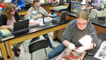
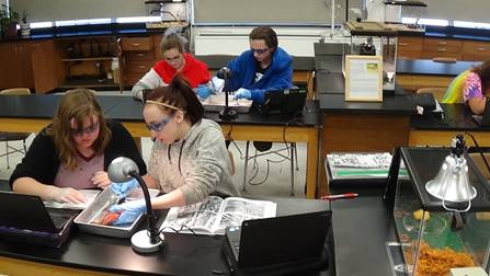
Method
Apparatii
|
Starfish Specimen: ie. Asteria or Pisaster
Dissection Tray with waxed bottom Razor Scalpel (blade #11)
Magnifying Lens (or Stereoscope/Compound) Forceps |
Scissors (fine point) Needle Probe Blunt Probe Gloves
|

Procedure: May need to refer to the “Directions and Planes in Anatomical Description Chart”
External Anatomy
1. Compare your specimen to figures and illustrations provided. Use these diagrams to familiarize yourself with the various external features of the animal.
Ray = Limb of a starfish.
Spines = Protrude from the surface of the endoskeleton and give the body a spiny appearance. Using a magnifying glass or scope, one should notice that there is a thin epidermal covering on top of the movable spines. Spines help in burrowing.
2
Central “Disc” = Where all rays are connected together collectively. Activity center of the starfish and contains the mouth.
Madreporite = Large button-like structure that is located off center on the Central Disc between two rays. It is the opening of the “water-vascular system”. The inconspicuous Anus is in the center of the disc and both structures are located on the upper, dorsal or aboral surface of the starfish.
Mouth = Center of the underside, ventral or oral surface of the starfish body.
Ambulacral Grove(s) = On the oral surface of each ray extending from the mouth to the tip of each ray. These deep furrows contain two or four rows of Tube Feet, with protruding Suckers.
Eyespot = A small whitish circle/bump found at the end of each ray (containing red pigment if a fresh specimen) which allows the starfish to sense and respond to light. It is best to use a magnifying glass or scope to see.
Internal Anatomy
2. Compare your specimen to the figures and illustrations provided. Use these diagrams to familiarize yourself with the various internal features of the animal. Concentrate primarily on the bold terms and try your best to find each structure listed.
3. Cut one ray off your specimen and study the cross section of the stump.
4. Make a cut up the sides of the severed Ray to allow you to open the body wall and view the structures and pattern of the inner skeletal framework.
5. Observe the following: Pyloric or Hepatic Caeca (digestive glands), Ambulacral Ridge (contains the radial canal), and the clear bubble-like Ampullae (small dilatation in a canal or duct). Remove the Caeca from a ray. If you have clearish / pink “tissues”, these are Gonads (sexual organs) that are under the Caeca, alongside the Ambulacral Groove.
6. Sever the “tip” from an undissected ray and then cut up each side of the ray towards the central disk and carefully remove the central Oral Wall.
7. Turn the specimen to the Aboral side. Carefully remove the middle aboral body wall, separating the organs from the internal surface of the body wall by clipping the Mesenteries, but leaving the Madreporite intact.
8. Find the stomachs which are referred to as the Pyloric and Cardiac Stomachs. The cardiac stomach is the large, clearish, bag-like structure which protrudes through the mouth while feeding. The cardiac stomach is retracted by five pairs of retractor muscles, one pair in each ray. Remove the stomachs in order to view structures of the “water-vascular system”.
9. Review and/or find the following structures that make up the “water-vascular system”: Madreporite, Stone Canal, Ring Canal, and Radial Canal (within or part of the “Ambulacral Ridge”)
10. There is a Hemal system (relating to the blood or blood vessels) present in starfish but difficult to find. Circulation of fluids is accomplished by the action of cilia that line the body cavity.
11. Clean and return all materials as instructed during class.