Contraction of the Heart Mini-Labs, 2018
Section #1: Pulse
What is the pulse rate?
The pulse rate is a measurement of the heart rate, or the number of times the heart beats per minute. As the heart pushes blood through the arteries, the arteries expand and contract with the flow of the blood. Taking a pulse not only measures the heart rate, but also can indicate the following:
- heart rhythm
- strength of the pulse
The normal pulse for healthy adult’s ranges from 60 to 80 beats per minute. The pulse rate may fluctuate and increase with exercise, illness, injury, and emotions. Females ages 12 and older, in general, tend to have faster heart rates than do males. Athletes, such as runners, who do a lot of cardiovascular conditioning, may have heart rates near 40 beats per minute and experience no problems.
How to check your pulse:
As the heart forces blood through the arteries, you feel the beats by firmly pressing on the arteries, which are located close to the surface of the skin at certain points of the body. The pulse can be found on the side of the lower neck, on the inside of the elbow, at the wrist or near your ankle. When taking a pulse:
1. Obtain a stopwatch or second hand
2. Using the first and second fingertips, press firmly on the arteries of your partner wrist until you feel a pulse.
3. Begin counting the pulse when the clock's second hand is on the 12.
4. Count your pulse for 60 seconds (or for 15 seconds and then multiply by 4 to calculate beats / minute).
5. When counting, do not watch the clock continuously, but concentrate on the beats of the pulse.
6. Have your partner record their pulse on their handout.
_____ / min. (your pulse rate)
7. Repeat steps (1-6) with your partner. Determine their pulse rate:
_____ / min. (your partner’s rate)
8. Have your partner jog / walk very quickly (back and from and around the gym) and repeat steps 1-7.
Both you and your partner are to exercise separately.
_____ / min. (your pulse rate after exercising)
9. Why would there be a difference in pulse rate after the exercise?
10. There are two sounds heard during each heart beat. These are called Lub-Dub noises by doctors. When the valves between the upper chambers (atria) and lower chambers (ventricles) close, a "lub" sound is heard. When the valves in the pulmonary and aortic arteries leaving the heart close, a "dub" sound is heard followed by a longer pause Lub-DubLub-Dub. Have your partner take the “bell” of a stethoscope and place it on their heart.
Did you hear the valves of their heart opening and closing? (Yes or No) ________
Extra:
1. Have your partner remove one of their shoes and socks.
2. Using your first and second fingertips, press firmly on the arteries just below your partner’s medial malleolus bone.
3. Feel for a peripheral pulse similar to what you would find anteriorly along the neck, elbow or wrist.


Section #2: Blood Pressure
What is blood pressure?
Blood pressure, measured with a blood pressure cuff and stethoscope by a nurse or other healthcare provider, is the force of the blood pushing against the artery walls. Each time the heart beats, it pumps blood into the arteries, resulting in the highest blood pressure as the heart contracts. One cannot take his/her own blood pressure unless an electronic blood pressure monitoring device is used. Electronic blood pressure monitors may also measure the heart rate, or pulse.
Two numbers are recorded when measuring blood pressure. The higher number, or systolic pressure, refers to the pressure inside the artery when the heart contracts and pumps blood through the body. The lower number, or diastolic pressure, refers to the pressure inside the artery when the heart is at rest and is filling with blood.
High blood pressure, or hypertension, directly increases the risk of coronary heart disease (heart attack) and stroke (brain attack). With high blood pressure, the arteries may have an increased resistance against the flow of blood, causing the heart to pump harder to circulate the blood.
The guidelines define normal blood pressure as follows:
- Less than 120 mm Hg systolic pressure
- Less than 80 mm Hg diastolic pressure
These numbers should be used as a guide only. A single elevated blood pressure measurement is not necessarily an indication of a problem. A person who normally runs a lower-than-usual blood pressure may be considered hypertensive with lower blood pressure measurements than 140/90. Keep in mind that certain factors can cause blood pressure to temporarily raise. Blood pressure normally rises as a result of:
- Stress
- Smoking
- Cold temperatures
- Exercise
- A full stomach
- Full bladder
- Caffeine
- Certain medicines
How to check your blood pressure:
1. Place the cuff on the upper “bare” arm of your partner.
2. Place the bulb in your hand and place the gauge where you can observe it.
3. Close the airflow valve on the bulb by turning the screw clockwise.
4. Firmly hold and place the “diaphragm” of the stethoscope just below the cuff and under the elbow.
5. Inflate the cuff by squeezing the bulb with your right hand several times. You may hear the pulse in the stethoscope.
6. Watch the gauge. Keep inflating the cuff until the gauge reads about 30 points (mm Hg) above your expected systolic pressure (~120 mm Hg). At this point, you should not hear the pulse in the stethoscope.
7. Keeping your eyes on the gauge, slowly release the pressure in the cuff by opening the airflow valve counter clockwise. The gauge should fall only 2 to 3 points with each heartbeat. (You may need to practice turning the valve slowly.)
8. Listen carefully for the first pulse beat. As soon as you hear it, note the reading on the gauge. This reading is your systolic pressure.
9. Continue to slowly deflate the cuff.
10. Listen carefully until the sound disappears. As soon as you can no longer hear your pulse beat, note the reading on the gauge. This reading is your diastolic pressure. Allow the cuff to completely deflate.
Record your blood pressure: ________________ (may wish to take a few times an average)


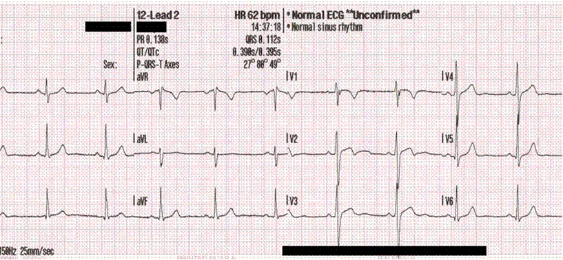
Section #3: EKG (or ECG) / Heart Monitor via Probe-ware
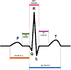
What does an EKG wave tell?
During normal atrial depolarization, the main electrical vector is directed from the SA node towards the AV node, and spreads from the right atrium to the left atrium. This turns into the P wave on the EKG
The QRS complex is a structure on the EKG that corresponds to the depolarization of the ventricles. Because the ventricles contain more muscle mass than the atria, the QRS complex is larger than the P wave.
The T wave represents the repolarization (or recovery) of the ventricles.
How to read the basic
components of an EKG:
(see diagram of a
“normal” EKG)
1. Note that each measurement represents a “lead: II, III, IIII” which simply means areas where the electrodes were placed on the patient.
2. Using a pencil/pen, neatly circle one (1) normal EKG “wave” (hint: P + QRS + T)
3. Using a pencil/pen, neatly label a P wave
4. Using a pencil/pen, neatly label a Q and then an R and then an S wave
5. Using a pencil/pen, neatly label a T wave
6. If one 5 mm x 5 mm block = .2 seconds (200 ms) of time, then approximately what is the interval of the (1) normal EKG “wave”? (Hint: measure from the beginning of the P wave to the end of the T wave)
______ seconds
7. If one 5 mm x 5 mm block = .5mV of amplitude, then approximately how many amps were recorded from the second EKG “wave” from lead III of a normal heart for the following waves?
P wave _____ mV ; the “R” in the QRS complex _____ mV ; T wave _____ mV
8. What is happening to the heart during a P wave?
9. What is happing to the heart during a QRS complex set of waves?
10. What is happing to the heart during a T wave?