Modeling DNA, RNA, and Protein Synthesis Activity 2018
Introduction
Traits are passed on from one generation to the next and are based on the types of proteins that are made by the cell. All organisms carry an elaborate blueprint containing the information necessary to develop and maintain life. This genetic instruction manual is located in a chemical molecule called DNA, which is found within a person’s genes (sections of a DNA molecule). The instructions are passed to mRNA, which provides a sequence of bases (code) for tRNA to bring amino acids to a ribosome where the two molecules meet. As a result, a “protein” is made.
|
|
|
General Protein Synthesis Simulation: “Translation”
Part 3
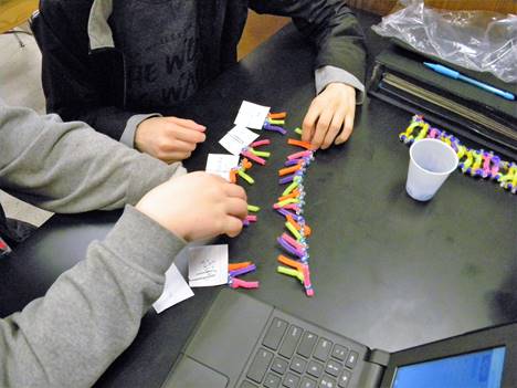
The ribosome in the cytoplasm is where Translation takes place. The ribosome binds to the mRNA at the start codon (AUG). The ribosome then goes along the mRNA strand. While the ribosome is moving, amino acids, linked to tRNA, bind to their appropriate codon on the mRNA; thus, forming complementary base pairs within the ribosome. As the ribosome continues to move from codon to codon along the mRNA, amino acids are added one by one by means of peptide bonds. At the end, the nonsense codon “stop codon” terminates translation and a protein is released.
Apparatii
Stock Table
purple chenille stems (pipe cleaner) = cytosine
pink chenille stems (pipe cleaner) = guanine
green chenille stems (pipe cleaner) = adenine
orange chenille stems (pipe cleaner) = uracil (note: yellow for thymine)
Group:
* Previously made DNA model
* Previously made mRNA model
(5) large paper clips = for tRNA backbone (phosphodiester bonds)
note card (large-standard/no lines preferred) = amino acids
tape = peptide bonds
Dixie-cup (to hold beads)
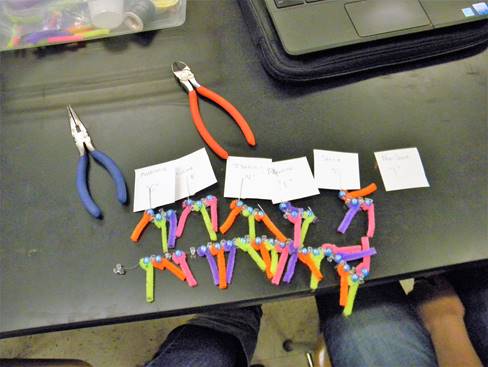
wire cutter
pliers
scissors
fine black marker
pencil
references
|
|
|
Procedure
1. Gather one (1) of each “chenille stem” (pipe cleaner) color [not for Thymine]. Using a ruler and wire cutter carefully and exactly cut the “chenille” stems into (6) 5 cm “base” pieces. (note: using a black marker to measure the stems prior to cutting may help)
2. With the “large” paper clips, bend down only one outer wire into a “right angle” leaving the rest of the clip alone to be able to hold a piece of paper.
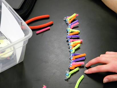
3. Gather (15) light blue Barrel Pony Beads and (10) Tri-Beads. Using your understanding of RNA and/or references, construct a single tRNA molecule by stringing first a Barrel Pony Bead to the wire and then stringing a Tri-Bead until you have strung three (3) Barrel Pony Beads and (2) Tri-Beads. Note that the rest of the paper clip wire is to simulate the continuation of the “sugar-phosphate backbone” of an RNA molecule; about 80 nucleotides long.
4. Using pliers, slightly bend the one end of each paper clip that was placed into a “right angle” upwards/around to prevent the beads from falling off. Complete 2-4, 4x’s in order to have a total of 5 tRNA’s
5. Using the just created tRNA molecules, attach the 5 cm “chenille” bases using the following key:
tRNA “anti-codon” Base Key
* Note: each tRNA molecule will only have three (3) bases attached to their sugars; collectively these bases are called “anti-codons”
|
purple = cytosine (C) pink = guanine (G) |
green = adenine (A) orange = uracil (U) |
|
tRNA #1: U C G |
tRNA #3: U A U |
tRNA #5: C G A |
|
tRNA #2: G A G |
tRNA #4: U A C |
|
· Only thread and bend around one end of the “chenille” base threw the “Barrel-Bead” sugar; the other end of the “chenille” base will be used later.
· Be sure to place the sequence from left-to-right for every tRNA molecule.
· The bases on the tRNA molecule are called, “anti-codons” and will be used to bond with complementary bases on the mRNA called, “codons”.
6. Using the note card, bend it into 6 equal squares (bend once horizontally and then 3x vertically). Using scissors, cut out the squares that you outlined by bending the card.
7. Using the table below, write the following names of these amino acids or function and letter in quotes (“”) on each “side” of each card (note: the letters in quotes (“”) are added to make sure you have the right sequence when you are done with this exercise; a hidden word, which means an area on a DNA that is copied to make mRNA, will form if the amino acids placed correctly):
|
for tRNA #1:Serine “S” |
for tRNA #3:Isoleucine “N” |
tRNA #5: Alanine “E” |
|
for tRNA #2:Leucine “E” |
for tRNA #4:Methionine “G” |
Non-Sense (stop) “!” |
|
|
|
8. Match each amino acid (card) with the correct tRNA by placing the amino acid (card) in the paper clip. For example, tRNA #1 needs to have “Serine” attached.
9. Create a “protein” by using the anti-codon bases on the tRNA molecules and the codon bases on the mRNA molecule. Match up each tRNA’s anticodon with its complementary codon on the mRNA molecule. You may want to use the chart below to confirm your amino acid -to- mRNA codon match. It is not necessary to attach the bases.
10.As you match the “complementary bases”, create a “peptide bond” between the amino acids by taping the cards (amino acids/functions) together AND releasing the “polypeptide” card from the tRNA (paper clip) molecule(s). Remember, the “Nonsense” card has no amino acid or tRNA associated with it; thus, it’s not part of the protein.